
Microscopy
We have invented a new type of two-photon microscope that can perform ultra-fast 3D random access imaging, enabling us to measure spatially distributed signaling in axons, dendritic trees and neural populations deep within brain tissue.
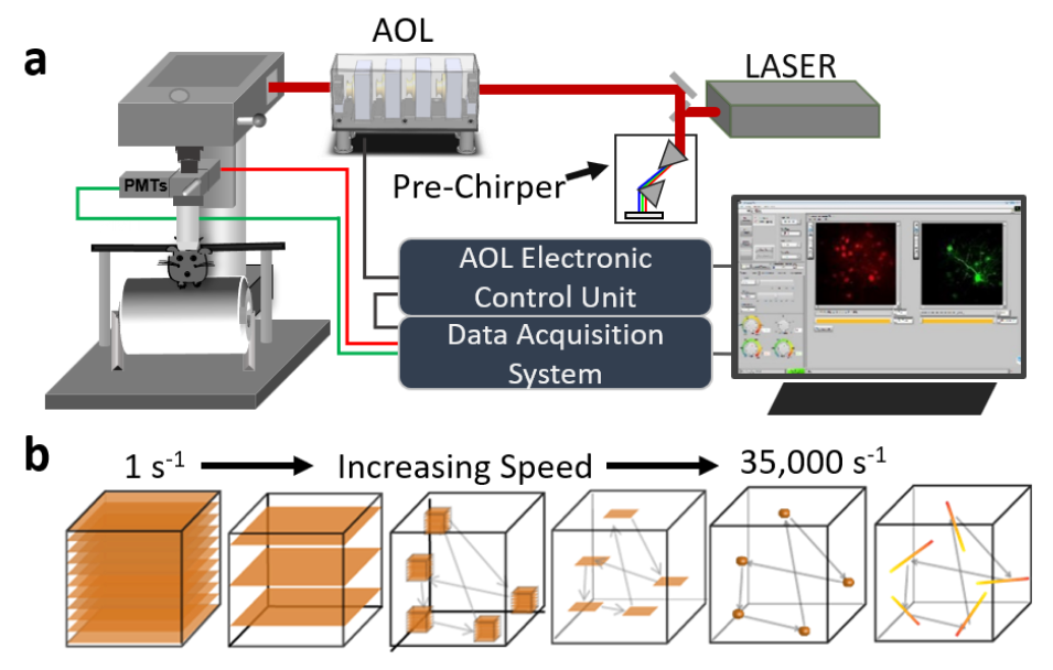
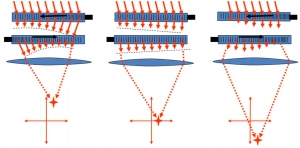
Schematic of AOL microscope with imaging modalities.
The Motivation
Our current understanding of how individual neurons and circuits represent and process information is limited by conventional microscope technology, which is relatively slow and restricted to 2D planes. This is problematic for measuring neural signaling events, since they are brief (1-100 ms), often occur in small structures such as synapses (~1 µm) and are spatially distributed in 3D space over large scales (0.1 – 1 mm). Moreover, the brains of awake behaving animals move. To meet these requirements, we have invented a compact Acousto-Optic Lens (AOL) 3D laser scanner (Kirkby et al., 2008, 2010, 2011, 2012) that performs high-speed multiscale 3D two photon imaging (Fernandez-Alfonso et al., 2014; Nadella et al. 2016) and corrects for brain movement in real time (Griffiths et al., 2020).
The compact Acousto-Optic Lens (AOL)
The high-speed 3D two-photon AOL laser scanner consists of 4 Acousto-Optic Deflectors arranged in series, driven with radio frequency ultrasound waves. The AOL deflects and focuses a two-photon laser beam to any arbitrary position within the imaging volume with high precision in the time that it takes a sound wave to travel across the aperture of the AODs (24.5 µs or ~ 40 kHz bandwidth). The AOL can generate a panoply of fast 3D imaging modes, from full frame raster scanning (30 Hz per frame for 512 x 512 pixel frames) over a 400 × 400 × 400 μm imaging volume to high speed continuous line scans in any orientation (20 kHz per line). Our AOL microscopes can track and correct for brain movements in real time using closed loop FPGA-based processing (Griffiths et al. 2020) and can operate at a range of wavelengths (800 nm – 1055 nm). The latest design of our AOL 3D scanner is compact (30 cm x 10 cm x 10 cm) enabling it to be fitted to existing two-photon microscopes by inserting the AOL into the optical path. Moreover, our microscope user software and analysis pipelines are open source (see Software Resources) The theory and implementation of AOL 3D scanning technology is described in (Kirkby et al., 2010; Evans et al., 2015; Konstantinou et al 2016; Nadella et al 2016; Griffiths et al. 2020) and has been patented by UCL Business (Kirkby et al., 2008, 2011, 2012).
Current applications
We are currently using AOL 3D two-photon microscopy to study how sensory and motor information is represented and transformed in cerebellar cortex and neocortex. To do this we have recorded activity from large populations of axons (Lanore et al. 2021) and inhibitory neurons (Gurnani and Silver, 2021) in the cerebellum and imaged the dendritic activity of neocortical pyramidal cells in awake animals as they perform behavioral tasks (see Neuroscience).
Microscope development
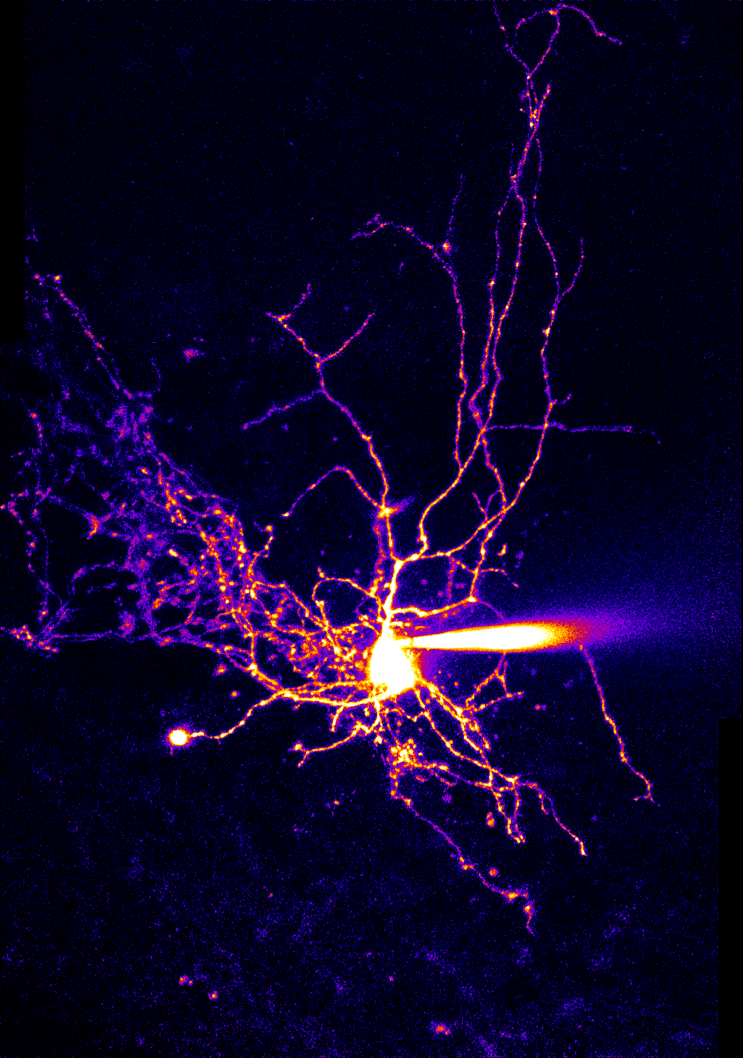
We continue to actively develop AOL 3D laser scanning technology to expand its functionality and improve its performance. Recent work includes compensation for linear and nonlinear field distortions enabling ultra-high precision (selective imaging of the entire dendritic tree of a neuron. By only imaging regions of interest much higher speeds can be achieved than for volumetric imaging. Our current work is focusing on improving the optical resolution of AOL microscopy, using AOL-based wavefront shaping and optimizing AOL 3D scanning for voltage imaging. In addition, we are actively disseminating our AOL 3D laser scanning technology to international collaborators, as part of a Brain Initiative (NIH) funded project.
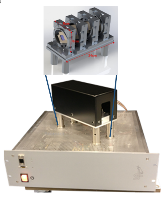
Latest design of AOL optical component and electronic control system.
Microscopy References
- Evans, G. J., Kirkby, P. A., Naga Srinivas, K., Marin, B. & Angus Silver, R. Development and application of a ray-based model of light propagation through a spherical acousto-optic lens. Opt. Express 23, 23493 (2015).
- Fernández-Alfonso, T. et al. Monitoring synaptic and neuronal activity in 3D with synthetic and genetic indicators using a compact acousto-optic lens two-photon microscope. J. Neurosci. Methods 222, 69–81 (2014).
- Griffiths, V. et al., Real-time 3D movement correction for two-photon imaging in behaving animals
Nature Methods, 17;741. [doi:10.1038/s41592-020-0851-7] - Kirkby, P. A., Srinivas Nadella, K. M. N. & Silver, R. A. A compact Acousto-Optic Lens for 2D and 3D femtosecond based 2-photon microscopy. Opt. Express 18, 13721–13745 (2010).
- Paul Kirkby, N.K.M. Naga Srinivas, R. Angus Silver. Methods and apparatus to control acousto-optic deflectors (WO/2012/143702) Inventors Dr https://patentscope.wipo.int/search/en/detail.jsf?docId=WO2012143702
- Paul Kirkby, N.K.M. Naga Srinivas, R. Angus Silver. Methods and apparatus to control acousto-optic deflectors (WO/2011/131933). Inventors Dr https://patentscope.wipo.int/search/en/detail.jsf?docId=WO2011131933
- Paul Kirkby, R. Angus Silver and N.K.M. Naga Srinivas. Imaging apparatus and methods (WO/2008/032061) Inventors https://patentscope.wipo.int/search/en/detail.jsf?docId=WO2008032061
- Konstantinou, G. et al. Dynamic wavefront shaping with an acousto-optic lens for laser scanning microscopy. Opt. Express 24, 6283–6299 (2016).
- Nadella, K. M. N. S. et al. Random-access scanning microscopy for 3D imaging in awake behaving animals. Nat. Methods 13, 1001–1004 (2016).
- Valera, A. et al., Precompensation of 3D field distortions in remote focus two-photon microscopy
Biomed. Opt. Express 12, 3717-3728 [doi:10.1364/BOE.425588]
Research supported by: